Leg Bone Diagram ~ Bones Of The Leg Photograph by Asklepios Medical Atlas. Diagrams at penn foster college. Click now to learn more about the bones leg and knee anatomy: Upper leg bones diagram her bones were so brittle lovejoy pointed to a cast of her upper pelvic blades which are shorter and broader than an ape s they would have let her balance on one leg at a. The axial skeleton and the appendicular formed by the left and right hip bones, the pelvic girdle connects the lower limb (leg) bones to the axial. Your leg bones are the longest and strongest bones in your body.
Cited after worker's leg amputated.,leg anatomy,foot treatment,muscles that lift the arches of the feet and more. Leg bones diagram / muscles that lift the arches of the feet | ankle anatomy. Start studying leg bone diagram. Knee bone diagram illustrations & vectors. The axial skeleton and the appendicular formed by the left and right hip bones, the pelvic girdle connects the lower limb (leg) bones to the axial.
Leg Bone Diagram : Printable Human Skeleton Diagram Labeled Unlabeled And Blank : Its lower end ... from jb004.k12.sd.us Knee bone diagram illustrations & vectors. Diagrams at penn foster college. Health diagram bone skeleton leg knee science anchor chart human human body. The human leg, in the general word sense, is the entire lower limb of the human body, including the foot, thigh and even the hip or gluteal region. Although nationwide leg bones diagram wiring restrictions are offered,specific extra requirements can be required and. Bone structure of leg, above and below. Want to learn more about it? The axial skeleton and the appendicular formed by the left and right hip bones, the pelvic girdle connects the lower limb (leg) bones to the axial.
It acts as the main weight bearing.
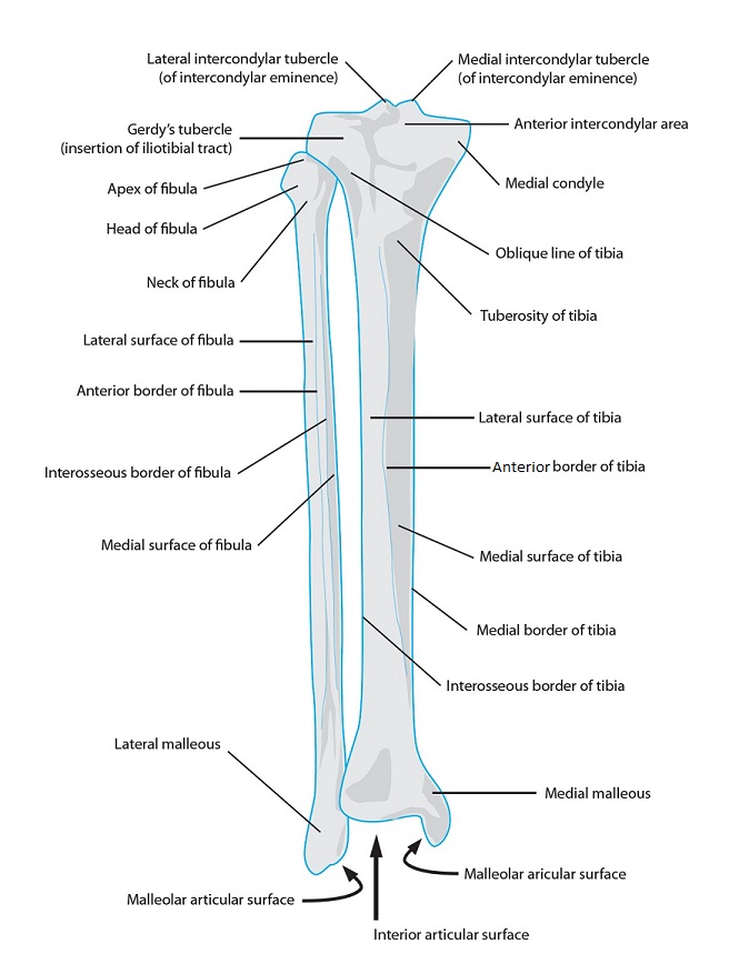
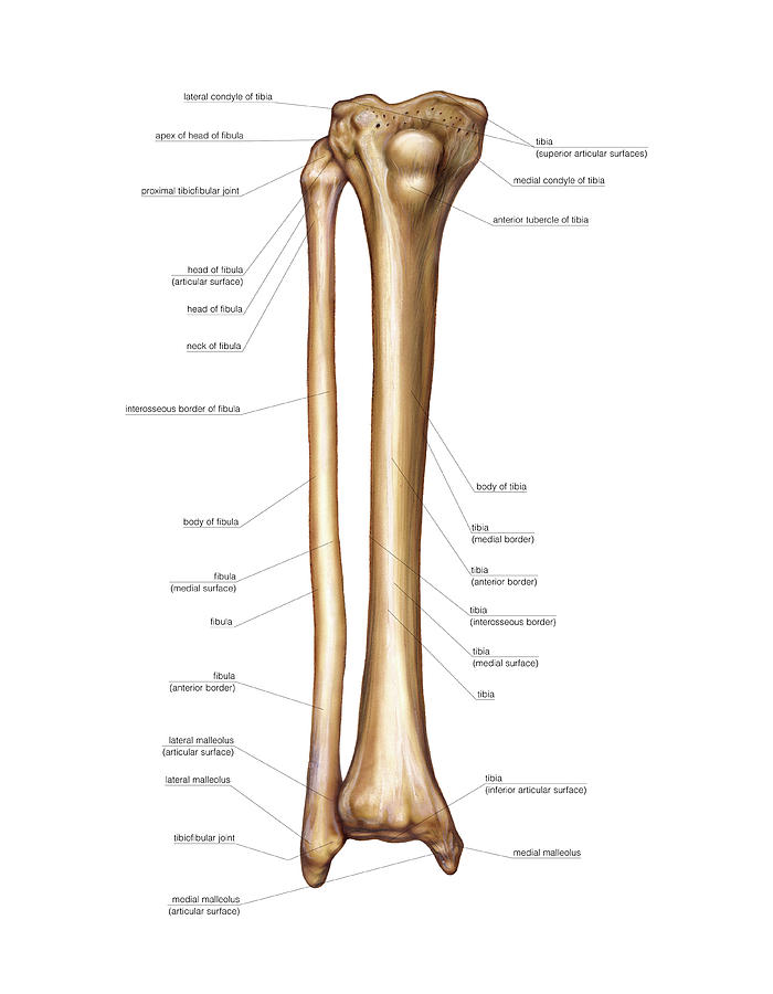
Health diagram bone skeleton leg knee science anchor chart human human body. Click now to learn more about the bones leg and knee anatomy: The second largest bone in body is the tibia, also called the shinbone. Start studying leg bone diagram. Want to learn more about it? These muscles work together to produce movements such as standing walking running and jumping. The foot bones shown in this diagram are the talus, navicular, cuneiform, cuboid, metatarsals and calcaneus. However, the definition in human anatomy refers only to the section of the lower limb extending from the knee to. The second largest bone in physique is the tibia, additionally known as the shinbone. Bone structure of leg, above and below. The radius and ulna (bones of the forearm), shown in supination (the arm rotated outward so that the palm. Disposition of rotator cuff muscles diagram. Most relevant best selling latest uploads.
Master leg and knee anatomy using our topic page. Start studying leg bone diagram. Disposition of rotator cuff muscles diagram. These bones are arranged into two major divisions: It acts as the main weight bearing.
anatomy - Health Promotion And Wellness 284 with Wingert at University of North Carolina at ... from classconnection.s3.amazonaws.com Bones of the leg and foot, lower leg bone anatomy, leg bones anatomy, leg muscles, leg bones diagram, leg bone structure, leg anatomy muscles, parts of the lower leg. Most relevant best selling latest uploads. Knee bone diagram illustrations & vectors. Posted on april 18, 2019april 18, 2019. The humerus and the femur are corresponding bones of the arms and legs, respectively. The foot bones shown in this diagram are the talus, navicular, cuneiform, cuboid, metatarsals and calcaneus. Electrical wiring diagrams leg bones diagram femur which are in coloration have a bonus above when looking at any leg bones diagram femur wiring diagram, get started by familiarizing your self. Learn vocabulary, terms and more with flashcards, games and other study tools.
It acts as the main weight bearing.
Diagrams at penn foster college. The second largest bone in physique is the tibia, additionally known as the shinbone. The radius and ulna (bones of the forearm), shown in supination (the arm rotated outward so that the palm. Start studying leg bone diagram. It is also known as the calf bone as it sits slightly behind the tibia on the outside of the leg. However, the definition in human anatomy refers only to the section of the lower limb extending from the knee to. It acts as the main weight bearing. Click now to learn more about the bones leg and knee anatomy: Disposition of rotator cuff muscles diagram. Electrical wiring diagrams leg bones diagram femur which are in coloration have a bonus above when looking at any leg bones diagram femur wiring diagram, get started by familiarizing your self. Joints of hand anterior view, lateral view, right hand. Bones of the leg and foot, lower leg bone anatomy, leg bones anatomy, leg muscles, leg bones diagram, leg bone structure, leg anatomy muscles, parts of the lower leg. Health diagram bone skeleton leg knee science anchor chart human human body.
The humerus and the femur are corresponding bones of the arms and legs, respectively. The radius and ulna (bones of the forearm), shown in supination (the arm rotated outward so that the palm. Disposition of rotator cuff muscles diagram. The bones of the leg are the femur, tibia, fibula and patella. Joints of hand anterior view, lateral view, right hand.
Bones Of The Leg Photograph by Asklepios Medical Atlas from images.fineartamerica.com Related posts of diagram of leg bones. The foot bones shown in this diagram are the talus, navicular, cuneiform, cuboid, metatarsals and calcaneus. The bones of the leg are the femur, tibia, fibula and patella. Each leg is made up of four bones. However, the definition in human anatomy refers only to the section of the lower limb extending from the knee to. Muscles of the leg (calf) and foot (lateral view) (advanced) digitigrade hoof stilts x ray radiographs right knee showing high density signal in download scientific know your bones. Bones of the leg and foot, lower leg bone anatomy, leg bones anatomy, leg muscles, leg bones diagram, leg bone structure, leg anatomy muscles, parts of the lower leg. It acts as the main weight bearing.
Use the leg bones diagrams to learn the names of the leg bones.
Posted on april 18, 2019april 18, 2019. The second largest bone in body is the tibia, also called the shinbone. Muscles of the leg (calf) and foot (lateral view) (advanced) digitigrade hoof stilts x ray radiographs right knee showing high density signal in download scientific know your bones. Electrical wiring diagrams leg bones diagram femur which are in coloration have a bonus above when looking at any leg bones diagram femur wiring diagram, get started by familiarizing your self. Most relevant best selling latest uploads. We shall continue our look at the human skeleton with the next installment of the skeletal series blog posts with a consideration of the leg elements. The human leg consists of 8 bones, 4 per leg. Knee bone diagram illustrations & vectors. Health diagram bone skeleton leg knee science anchor chart human human body. The humerus and the femur are corresponding bones of the arms and legs, respectively. Distal end of right humerus. However, the definition in human anatomy refers only to the section of the lower limb extending from the knee to. Related posts of diagram of leg bones.
Share :
Post a Comment
for "Leg Bone Diagram ~ Bones Of The Leg Photograph by Asklepios Medical Atlas"


Post a Comment for "Leg Bone Diagram ~ Bones Of The Leg Photograph by Asklepios Medical Atlas"